Clinically, imaging patients with arrhythmias is challenging and can lead to non-diagnostic images. Our group has developed several different approaches to assess patients with irregular heart rhythms which have improved our ability to understand this irregular rhythm and improved image quality (decreasing the risk of a non-diagnostic study).
Real-Time Cardiac MRI
We have developed real-time cardiac magentic resonance imaging techniques to overcome the conventional sampling limitations of MRI systems. While the acquisition has far less data available, parallel imaging, non-cartesian k-space sampling and iterative reconstruction result in images which allow clear visualization of irregular heart rhythms. Below, we highlight how this enables improved evaluation of patients with arrhtymias. We can visualize each heartbeat enabling assessment of different beat types which occur during arrhtymia. In addition to visualization, we can perform quantification of ventricular function.
Real-time MRI of a patient with sinus rhythm followed by an arrhythmia
Recent publications include:
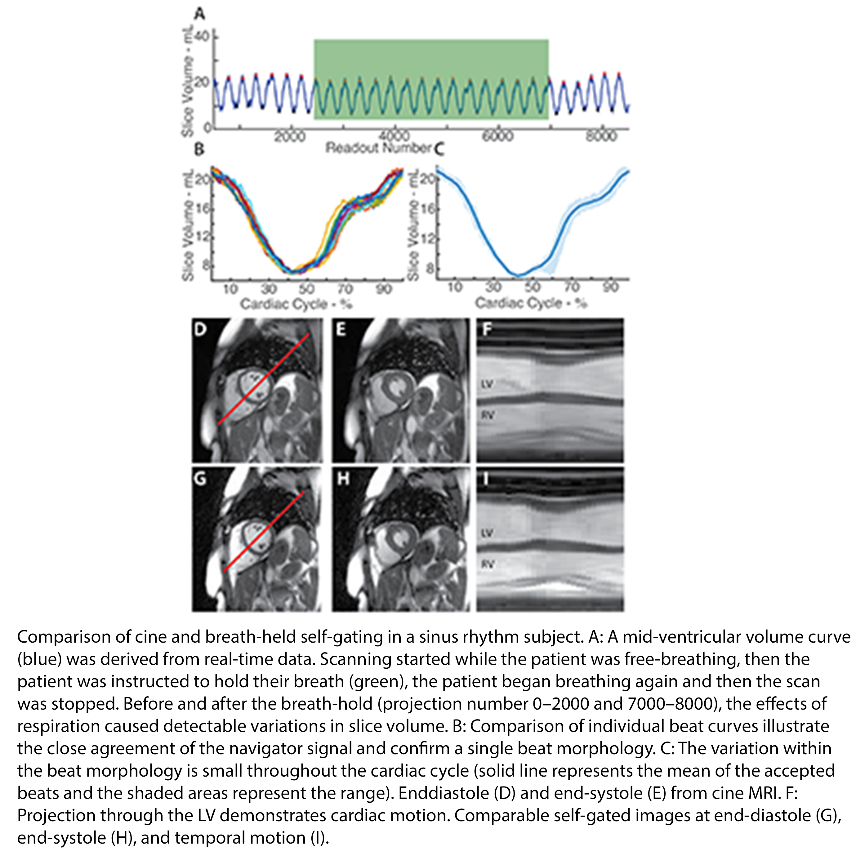
Self-gated MR Imaging
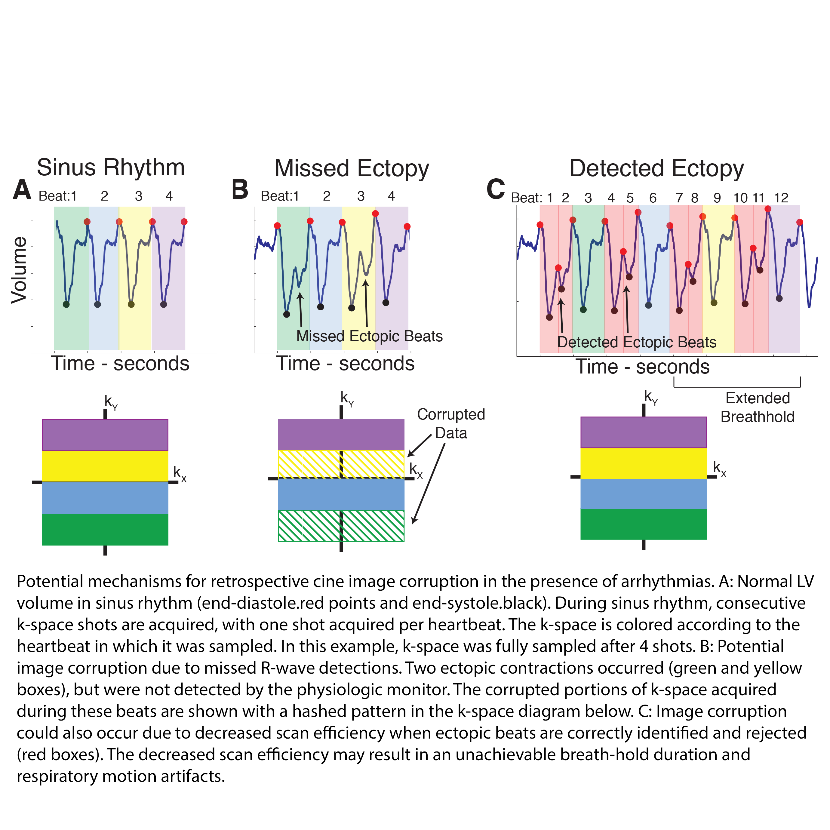
Conventional cardiac MR imaging techniques combine data acquired several heartbeats to increase image spatial and temporal resolution. This can prove challenging in patients with irregular heart rhythms. We have developed improved ways to sort imaging data based on physiologic information and provide high resolution imaging of arrhythmic beats
Recent publications include: