Our group has utilized the ability to image the heart during the entire cardiac cycle to characterize regional differences in regional right and left ventricular function.
We've also worked to both evaluate and improve the effectiveness of using CT to evaluate the presence of coronary disease.
Regional Estimates of Cardiac Function using cineCT
Modern CT scanners provide the opportunity to acquire low-dose images throughout the cardiac cycle. This allows for visualization of cardiac function in either ventricle. We have combined this acquisition with a quantitative assessment tool (SQUEEZ) to characterize the regional heterogenity of function.
Both ventricles can be visualized throughout the cardiac cycle
Regional estimates of RV strain in an adult patient with repaired Tetralogy of Fallot
Recent publications include:
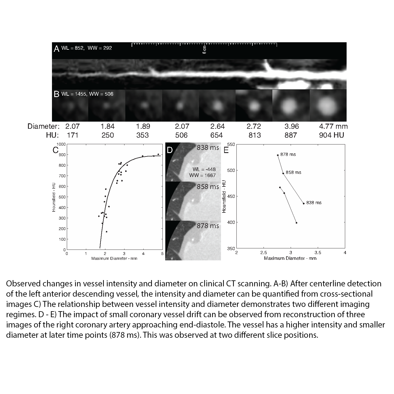
Coronary Artery Disease Imaging with CT
Modern CT scanners can be utilized to non-invasively detect coronary artery disease via coronary artery angiography. However, despite engineering efforts to accelerate data acquisition, there are small motions that can occur during imaging. We have utilized computer simulations to evaluate trade-offs inherent to conventional scanners in terms of spatial resolution and the sensitivity to motion.
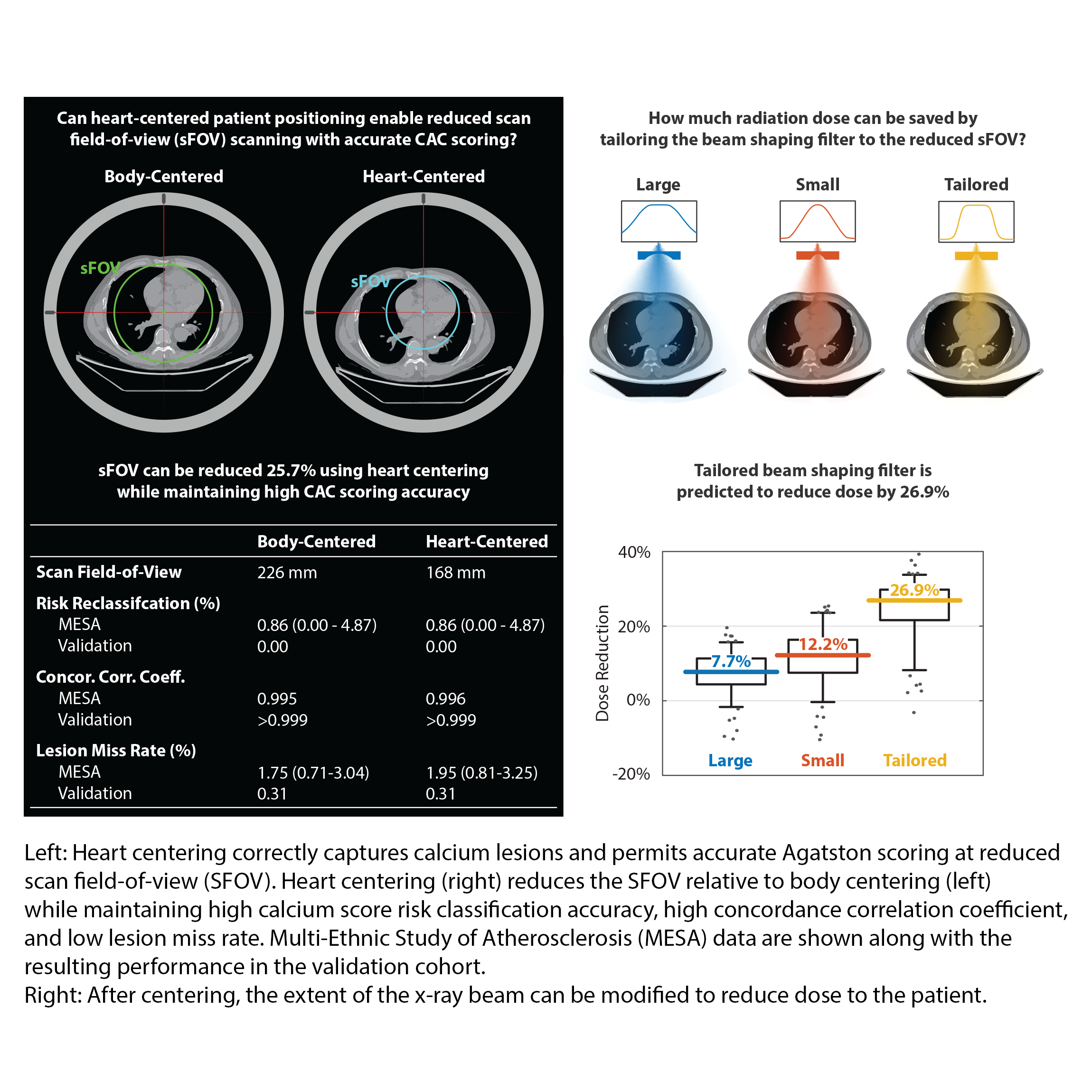
In addition, while the focus of these scans is the coronary vessels, the entire chest is exposed to radiation. We developed a model to improve patient positioning and showed that it can reduce dose to regions around the heart while maintaining diagnostic accuracy.
Lastly, we have develop genetic-based approaches to improve which patients are referred for imaging.
Recent publications include: